
Tick or Skin Tag on a Dog? Visible Clues and When to Ask a Vet
Learn how to tell a tick or skin tag on dog from photos and hands-on checks, which clues matter, and when to contact a vet for safe next steps.
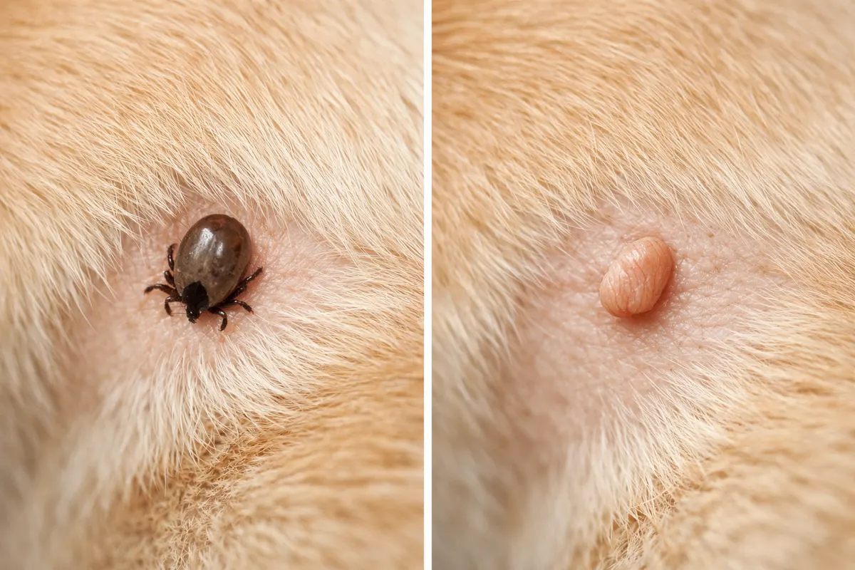
Quick answer: tell a tick from a skin tag on a dog
If you’re asking “tick or skin tag on dog?” start by looking for a distinct body, legs, or a small hard head: ticks usually have a rounded body and may show legs at the edge when viewed closely. Skin tags are soft, fleshy growths attached by a narrow stalk and move with the skin.
A photo that clearly shows scale (a ruler or a common object) and multiple angles—top, side, and a close-up—often reveals whether the bump has legs or a hard shell. If the bump is dark and shiny and you can see tiny legs or a mouthpart, treat it as a tick until proven otherwise.
When in doubt, don’t tug. If removal is necessary, use proper tick-removal technique or contact your veterinarian. A photo-based check can help you decide whether to remove immediately, watch for change, or seek veterinary care.
- Look for legs, mouthparts, or a segmented body — signs of a tick.
- Fleshy, soft, dangling lumps that move with the skin usually indicate a skin tag.
- If the lesion bleeds easily, is rapidly changing, or the dog seems unwell, call a vet.
What “tick or skin tag on dog” usually refers to
Owners use the phrase “tick or skin tag on dog” when they find a small, raised lesion and aren’t sure whether it’s a blood-feeding parasite or a benign skin growth. The distinction matters: a tick can attach, feed, and transmit disease; a skin tag is usually harmless but can irritate or be mistaken for other lesions.
Ticks vary in size and appearance across life stages. Larvae are tiny and hard to see, nymphs are small and harder to identify by eye, while adult ticks can be several millimeters across and more obvious. Ticks may appear flat before feeding and engorged and rounded after feeding.
Skin tags (acrochordons) are benign flaps of skin composed of the same tissue as surrounding skin. They often form in areas of friction or folds and typically don’t have a hard shell, legs, or a mouthpart. They tend to stay the same size or grow slowly.
Knowing this context helps you decide how urgently to act. If the lump is a tick, prompt removal reduces disease risk. If it’s a skin tag, unnecessary aggressive removal can harm the dog. A careful visual and tactile check helps prioritize your next steps.
- Ticks: external parasites with legs, mouthparts, and a hard or leathery body; may engorge after feeding.
- Skin tags: soft, flesh-like, often attached by a narrow stalk and usually motionless relative to the skin.
- Other lookalikes: scabs, cysts, warts, or embedded foxtail awns; context and behavior of the lump guide identification.
Key visual and tactile clues to check
Use these clues in combination. No single feature proves identity in every case, but a cluster of consistent observations raises confidence. Take photos from multiple angles, include a size reference, and gently note how the lump moves with the skin.
Color and texture: ticks are often uniformly dark brown, black, or gray when unfed and may become lighter or bloated after feeding. Skin tags match surrounding skin tone or are slightly darker and have a softer, wrinkled surface.
Shape and attachment: a tick has a rounded, sometimes segmented-looking body and appears attached by a small head embedded in the skin. A skin tag usually hangs by a slender stalk or sits flush with the skin without a defined separate head.
Movement and response: gently move the skin around the lump. A tick will remain attached in place; the body won’t slip the way a loose scab or a flake of dirt would. A skin tag will move with the skin and often bends at its stalk. Also check whether the dog reacts to pressure around the area.
- Size with scale: measure or place a coin/pen nearby in photos to record size accurately.
- Visible legs or mouthparts at the edge of the bump strongly indicate a tick.
- Shiny, hard shell or leathery surface is typical for ticks; soft, fleshy, or wrinkled texture suggests a skin tag.
- Look for a central dark point (mouthpart) at the skin surface — ticks use this to anchor attachment.
- If the lesion is mobile beneath the skin rather than attached at the surface, it may be a cyst or embedded foreign object, not a tick or skin tag.
Step-by-step workflow
Follow a calm, consistent routine to inspect any suspicious lump. Start by restraining your dog safely—ask a helper to hold them if needed—so you can examine without startling the animal. Keep lighting bright and steady for photos and inspection.
Document before you touch. Take at least three clear photos: a close-up from above, a side view to show profile and attachment, and a wider shot showing location on the body and an object for scale. Record when you first noticed the lump and any recent outdoor exposure.
Gently part the hair and use a magnifier or phone camera zoom to look for legs or a mouthpart. Avoid squeezing or attempting to rip the lump off. If you suspect a tick and removal is necessary, use fine-tipped tweezers or a tick tool, grasping the tick close to the skin and pulling steadily without twisting.
After removal, disinfect the bite site, save the specimen in a sealed container with a note of date and location, and monitor the dog for signs of illness (lethargy, fever, loss of appetite, lameness). If the lump resists removal, bleeds excessively, or the dog shows systemic symptoms, seek veterinary care promptly.
- Stage 1 — Observe: photograph with scale, note location, size, and behavior of the lump.
- Stage 2 — Inspect: part hair, use light/magnifier, look for legs, shell, mouthparts, or stalk.
- Stage 3 — Decide: if you see legs or a mouthpart, plan safe removal; if fleshy and stalk-like, consider monitoring or vet assessment.
- Stage 4 — After action: clean the site, save photos and specimen if removed, and record the removal time for vet follow-up if needed.
Examples
Example 1 — Tiny dark dot behind the ear: a small, dark, flat spot with no visible legs in a long-haired dog. After parting the fur, you see a tiny round body with faint legs at the edge when zoomed. This pattern fits an early-stage tick; removal with tweezers is reasonable and immediate observation for symptoms follows.
Example 2 — Soft tag on the belly fold: a small, pink, soft growth attached by a narrow stalk where skin rubs together. It doesn’t show hard shell or legs and bends when the skin moves. This looks like a skin tag; unless it causes irritation, schedule a vet check or monitor for change.
Example 3 — Raised scab-looking bump that flakes: a scab from a healing wound or insect bite can mimic a tick. Scabs are irregular, may flake, and aren’t uniformly rounded. Clean the area, avoid picking, and photograph for reference. If the lesion persists or grows, veterinary evaluation is appropriate.
Example 4 — Embedded seed or foreign body: sometimes a grass awn or foxtail sits under the skin and produces a small lump. It can cause local inflammation and may mimic a cyst. These often feel tense, may be painful to the touch, and usually require veterinary removal.
- Small dark rounded bump + visible legs → treat as tick and remove safely.
- Dangling, soft stalk-like lump → likely skin tag; monitor or consult vet if irritated.
- Irregular scab or crust → likely healing wound; don’t pick, watch for infection.
- Firm, painful lump under the skin → possible foreign body or abscess; seek a vet.
Limitations
Photos and home checks are powerful first steps but have clear limits. A photo cannot rule out tick-borne disease, determine whether a skin growth is malignant, or always identify very small or partly embedded ticks. Visual clues only guide next steps; they do not replace professional veterinary diagnosis.
Some lookalikes — like early cysts, small warts, embedded foxtail awns, or certain parasitic larvae — can mimic ticks or skin tags in appearance. Lighting, camera blur, hair, and swelling can hide critical detail such as tiny legs or a mouthpart. That’s why multiple angles, scale, and a gentle tactile exam matter.
Risk-based outcomes: if the lesion is clearly a tick with visible legs, removal is usually safe at home with proper technique, but you cannot assess infection risk from appearance alone. If the lump bleeds, resists removal, is rapidly changing, or the dog shows systemic signs, a veterinarian should evaluate and, where indicated, run diagnostic tests.
When to escalate: persistent lumps, growing masses, recurring irritation, or any signs of illness (fever, lameness, loss of appetite, unusual lethargy) require veterinary attention. Keep clear records of photos, removal time, and any clinical signs to share with the vet.
- A photo rarely proves absence of disease; testing may still be needed after a tick bite.
- Do not try forceful removal of suspicious lumps that don’t come free or cause heavy bleeding.
- If you’re unsure, capture good photos, monitor for change, and consult a vet rather than guessing.
- Avoid home remedies or harsh chemical treatments on lesions; they can worsen infection or hide symptoms.
Related guides
Want a guided first pass after your visual check?
If you’ve photographed a suspicious lump or completed the checks above, consider using the Dog Tick Check workflow at https://tick-detector.app/tools/dog-tick-detector as a structured next step. It’s designed to help you organize photos, record scale and location details, and flag the clues that matter most before you decide whether to remove or seek veterinary care. Use it as an evidence-gathering aid—not a diagnosis—and share your findings with your veterinarian when in doubt.
Take the next step →Frequently asked questions
How can I safely remove a tick from my dog at home?
If you decide to remove a tick yourself, use fine-tipped tweezers or a purpose-designed tick tool. Grasp the tick as close to the dog’s skin as possible—near the mouthparts—and pull straight outward with steady, even pressure. Avoid twisting, squeezing, or crushing the tick’s body to reduce the chance of pushing infectious fluids into the bite. After removal, clean the site with antiseptic, save a photo or the specimen in a sealed container, and note the time and location of removal. If the tick’s mouthparts remain embedded, or if the bite bleeds excessively, contact your veterinarian for assistance.
Can a photo alone confirm whether a lump is a tick or skin tag?
A clear, well-lit photo with scale and multiple angles can often identify visible features like legs, a hard shell, or a stalk, which strongly suggests a tick or skin tag. However, photos can miss tiny nymphs, partly embedded mouthparts, or internal characteristics of growths. Images can guide whether to remove, monitor, or seek a vet, but they cannot rule out infection, internal lesions, or microscopic causes—so follow-up may still be necessary.
When should I take my dog to the vet for a suspicious lump?
Seek veterinary care if the lump is rapidly growing, painful, bleeding, does not come off with careful removal, or if your dog shows systemic symptoms like fever, lethargy, lack of appetite, or lameness. Also consult a vet for lumps that recur in the same spot, change color or shape, or if you’re unsure after taking photos and attempting a careful inspection. A vet can perform tests, remove the lesion safely, and advise on tick-borne disease prevention and monitoring.
If I remove a tick, how long should I watch my dog for symptoms?
Monitor your dog for at least 30 days after removal for signs such as fever, decreased appetite, unusual tiredness, stiffness, limping, or swollen joints—these can indicate tick-borne disease. Keep a record of the removal date, tick appearance (if photographed or saved), and any new symptoms. If any concerning signs appear, visit your veterinarian promptly for evaluation and possible testing. Early veterinary evaluation improves management of tick-borne infections.