
Tick Lookalikes: Visual Clues, Clear Checks, and Safe Next Steps
Learn how to spot tick lookalikes with clear visual clues, when a photo is enough, and safe next steps. Practical checks before verification or vet help.
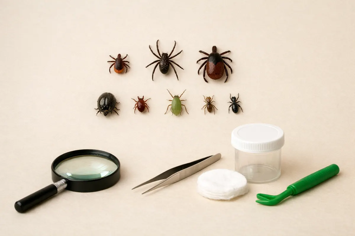
Quick answer: can this be a tick or a lookalike?
Most quick identifications rely on a few visible facts: body shape, leg count, and where the object is attached. If you see a rounded, flattened body with no visible antennae and a mouthpart at the front, it may be a tick; if the object has a segmented body with clear head, thorax and abdomen, antennae, or wing stubs, it is more likely an insect or mite lookalike.
Life stage matters. Larval ticks have six legs and can be mistaken for small insects; nymphs and adults have eight legs (they are arachnids). A tiny six-legged speck could be either a larval tick or a mite; resolving that requires better magnification or context.
If the object is attached to skin or embedded in hair with a visible feeding mouthpart (capitulum) or engorgement, treat it as a potential tick for removal and monitoring—do the safe removal steps and preserve the specimen if you need confirmation. If the object moves quickly, flies away, or has obvious insect features, it’s not a tick and usually needs no medical action.
Short takeaway: use leg count, body shape, mouthpart presence, and attachment behavior to triage. These clues reduce uncertainty but rarely prove species from one phone photo alone.
- Leg count: 6 legs can be larva or insect; 8 legs indicates nymph/adult arachnid.
- Attachment: a firm, biting attachment is more consistent with ticks; loose resting insects are usually not ticks.
- Body plan: ticks lack antennae and obvious segmented bodies seen in insects.
Tick lookalikes identification guide: definition and context
A tick lookalike is any small arthropod or object that can be mistaken for a tick in casual observation or a quick photo. That includes tiny insects (like beetle larvae and flies), mites, seed pods, scabs, grit, and plant debris. The term covers both true biological lookalikes and non-living items that mimic a tick’s shape or color.
For everyday users the practical question is not the taxonomy but the immediate risk and next step: does this need removal, monitoring, or no action? This guide frames how to decide by comparing visible anatomy (legs, mouthparts), behavior (attached and feeding versus crawling/flying), and context (where it was found and what the photo shows).
Life stage and scale complicate things. Tick larvae have six legs and can be extremely small; they are often mistaken for tiny insects. Nymphs and adults have eight legs and a more rounded body. A single photo with poor lighting or no scale bar frequently makes life-stage assignment impossible—so this guide emphasizes a checklist of visible clues and safe next steps rather than definitive species IDs.
Using the identification guidance here will help you sort sightings into high-confidence, probable, and uncertain categories so you can choose safe, proportionate actions: remove and preserve, document and monitor, or ignore and move on.
- Common non-tick lookalikes include mites, small beetles, lice, seed hairs, and skin scabs.
- Context matters: pets in tall grass or people handling brush are higher-probability tick encounters.
- If you need species-level ID (for research or medical reasons), plan to collect a preserved specimen or seek expert microscopy.
Key visual clues to spot tick lookalikes
Start with three quick visual checks: leg count, body segmentation, and presence of antennae. Under a macro photo or magnifier, count legs: six legs suggest either an insect or a tick larva; eight legs point to a nymph or adult tick or to mites (which are also arachnids). Because larval ticks have six legs, leg count alone is not definitive—pair it with attachment and body shape.
Examine body segmentation and shape. Ticks have a compact, broadly oval body without a distinct head/thorax/abdomen separation visible to the naked eye. Insects usually show a distinct head and thorax, and many have visible antennae or wing pads. Beetle or fly larvae are elongated or grub-like rather than the flattened, shielded shape of many ticks.
Look for the mouthparts and attachment. Ticks that are attached often show a small dark mouthpart (capitulum) at one end and will be immobile as they feed. Non-attached insects or debris move when prodded or will crawl away. Mites can be attached but are usually much smaller and often have different body textures under magnification.
Photo quality and scale are key contextual clues. A clear macro photo that includes a ruler, a coin, or a millimeter reference makes identification far easier. Also note where you found the object—on clothing, embedded in hair, on plant material, or on skin—because ticks are more likely to be attached in hair-bearing areas and in habitats like tall grass or leaf litter.
- Legs: count visible legs; if you only see six, consider larval tick vs insect and get closer images.
- Antennae: visible antennae = insect, not a tick.
- Segmentation: distinct head/thorax/abdomen suggests insect.
- Mouthparts: a forward-facing capitulum suggests a tick; insects won’t have the same structure.
- Movement: quick movement or flight rules out ticks.
- Texture: hard shield (scutum) or leathery cuticle can indicate certain tick families.
- Context clues: habitat, host (pet, human, clothing), and how it’s attached.
Step-by-step workflow to check a suspected tick lookalike
Follow a short, logical workflow that prioritizes safety and reduces uncertainty. First, pause and don gloves if the specimen is on a person or pet to avoid accidental exposure. If the object is freely moving, gently coax it into a clear container with a paper slip for observation. If it appears attached, focus on safe removal procedures rather than aggressive handling.
Next, document the sighting systematically. Take at least three photos: a close macro shot of the body, a side view that might show mouthparts, and a contextual photo showing where it was found (skin, hair, clothing, or plant surface) with a size reference. Record the date, location, recent outdoor activities, and whether the host has traveled to areas with known tick activity.
If removal is required, use fine-tipped tweezers or a dedicated removal tool. Grasp the specimen as close to the skin as possible and pull steadily without twisting. After removal, clean the area and preserve the specimen in a sealed container or between two pieces of tape if you may need verification. If the object detaches easily or flies away, it was likely not a tick and can be released outdoors.
Finally, triage your confidence. High confidence (clear eight-legged arachnid with visible bite): preserve specimen and monitor for symptoms. Probable but uncertain (tiny speck, poor lighting, or six legs): take additional photos, re-check with magnification, and consider asking a vet or local extension service for help. Low confidence (insect features, antennae, movement): no special action beyond routine hygiene.
- Step 1: Protect—wear gloves, don’t crush the specimen on skin.
- Step 2: Document—three photos: macro, side/mouthparts, and context with scale.
- Step 3: Remove—steady, close-to-skin grip; do not twist or squeeze.
- Step 4: Preserve—sealed container or tape for later ID if needed.
- Step 5: Triage—monitor or seek verification based on confidence level.
Examples: common situations and how clues change the conclusion
Example 1 — On a dog after a walk in tall grass: You find a small, dark speck embedded near the shoulder that won’t move. A macro photo shows a rounded body and what may be a mouthpart. This context and attachment behavior increase the likelihood it’s a nymph or adult tick. Safe next steps: carefully remove, preserve the specimen in a sealed container, and monitor the dog for signs of irritation or illness. If species-level ID or testing is needed, consult your veterinarian.
Example 2 — On clothing after gardening: A tiny bead-like object on fabric that moves when prodded and has visible antennae in a close-up photo is probably a small beetle or fly, not a tick. No removal from skin is needed; brush it off and check clothing seams. The presence of antennae and segmented head is the deciding clue here.
Example 3 — On a child’s arm that appears as a dark dot: A close macro image reveals a crab-like shape with eight legs and a flat body but no clear capitulum. If the object appears stuck, treat it as a tick for removal and preservation, but note that very small mites or chiggers can look similar. Because of the child's exposure and attachment, prioritize safe removal and, if the child develops a rash or symptoms, seek medical advice.
Example 4 — On a leaf or twig: Many plant seeds and bud scars look like small ticks. If the object detaches easily and has an irregular shape or fibrous texture under magnification, it’s plant debris. No animal or medical follow-up is needed; document the find only if you plan to report a suspicious species to local entomology resources.
- Dog in tall grass: attachment + rounded body = treat as tick and preserve.
- Clothing with antennae visible: insect, usually no action.
- Child with stuck speck: remove and monitor; consult medical advice for symptoms.
- Leaf/twig speck that detaches easily: plant debris, no medical action.
Limitations
A single photo, especially without scale or multiple angles, rarely supports a definitive species ID. Lighting, camera macro quality, and lens distortion can hide or exaggerate key features like mouthparts, scutum markings, or subtle segmentation. Expect a best-case outcome of a probable class (tick versus insect) rather than a safe species-level identification.
Life stage creates ambiguity: larval ticks (six legs) look like tiny insects; nymphs and adults (eight legs) may be mistaken for mites or other arachnids at small sizes. Even experienced identifiers often ask for multiple photos at different magnifications and a size reference before assigning a confidence level.
Do not infer disease presence or infectious risk from a photo. Pathogen carriage cannot be seen visually. Likewise, do not assume that an object is harmless because it looks like plant debris—attachment and behavior are the better clues to urgency.
When to escalate: if the specimen was attached and feeding, if the host develops unexplained symptoms, or if you need species-level confirmation for medical, public-health, or research reasons, preserve the specimen and consult a qualified lab, entomologist, public health authority, or your veterinarian. For pets, your veterinarian is the right first contact for both removal guidance and possible testing.
- Single-photo IDs are often provisional—expect uncertainty.
- Never use appearance alone to assess disease risk.
- Preserve specimens when attachment or symptoms occur.
- Seek expert or lab verification for species-level questions.
Related guides
Next step: check clues, then consult Tick Detector - Tickzen
After you’ve checked the visible clues above and preserved any attached specimen, use the Tick Detector - Tickzen identification chart to compare your notes and images. Treat the tool as a structured first pass to organize evidence—not a final diagnosis—and bring preserved specimens or clear photos to a vet, entomologist, or public-health lab for confirmation when needed. See the tick ID chart: https://tick-detector.app/tools/tick-identification-chart
Take the next step →Frequently asked questions
What should I compare when I spot tick lookalikes lookalikes on my dog's skin?
Compare leg count, body outline, and attachment. Check closely for antennae (which indicate insects) and count legs—six may be a larva or insect, eight suggests nymph/adult tick or mite. Note whether the object is embedded with a visible mouthpart. If it’s stuck and you can’t identify it confidently, remove it safely, preserve the specimen, and consult your veterinarian if the dog shows irritation or illness.
How can I tell a tick from a mite or small insect in a phone photo?
Zoom in on the body plan: ticks and mites are arachnids and typically show an oval, compact body; insects display distinct head, thorax, and abdomen and often have antennae. Look for eight legs (nymph/adult ticks and mites) vs six legs (insects and tick larvae). Also inspect for a mouthpart at the front and whether the specimen is attached and immobile. Because phone photos can hide details, take multiple angles and include a size reference to improve confidence.
Can a single photo confirm the tick species or disease risk?
No. A single image rarely confirms species and cannot indicate pathogen carriage. Visual features may suggest a genus in clear, high-resolution multi-angle photos, but species-level confirmation typically requires microscopic inspection or molecular tests performed by a lab. Treat visual IDs as triage: remove and preserve, monitor for symptoms, and seek expert verification if species-level ID or testing is important.
When should I seek medical or veterinary attention after finding a tick-like object?
Seek medical or veterinary attention if the specimen was attached and feeding, if the host develops a rash, fever, unexplained fatigue, or localized swelling, or if you are unsure whether removal was complete. For pets, contact your veterinarian for removal technique, preservation, and advice on testing and symptom monitoring. For people, contact a healthcare provider if symptoms develop; bring preserved specimen or photos to help clinicians assess exposure risk.