
Tick or Scab on Dog: What to Observe Before Touching
Quick, practical steps to decide whether a raised bump on your dog is a tick or a scab. Visual clues, safe checks, and when to contact your veterinarian.
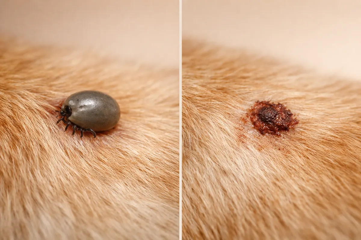
Quick answer: checking a bump for tick or scab on dog
When you spot a small dark bump on your dog's skin, the first priority is to observe without pulling. A tick will usually appear as a rounded, often slightly shiny body that may expand after feeding; a scab is typically flatter, crusted, and part of the surrounding skin. Neither a single photo nor a brief look is guaranteed—these visual cues only narrow possibilities.
If the bump has a visible set of legs or a distinct body outline that looks separate from the skin, treat it as a possible tick and follow safe removal guidance or seek veterinary help. If the lesion is crusted, scaly, or clearly attached by wider tissue rather than a discrete body, consider scab, healing wound, or skin growth until a professional confirms otherwise.
Record where you found it, take steady photos from several angles, and monitor changes over 24–48 hours. Keep handling minimal until you can determine whether it’s an attached arthropod (tick) or a skin lesion that requires veterinary assessment.
- A tick may look rounded and sit on the surface; a scab is usually part of the skin and more irregular.
- Visible legs or a mouthpart at the skin edge strongly suggest an attached tick.
- Avoid tugging at an uncertain bump; gather photos and notes first.
What it means when you wonder 'tick or scab on dog'
The question 'tick or scab on dog' reflects two very different causes that need different actions. A tick is an arthropod that attaches with a small mouthpart and feeds on blood; removing it promptly and properly reduces the chance of disease transmission and skin irritation. A scab is a sign the skin has already been injured or infected and is part of the body's healing response—this requires wound care or veterinary attention if it looks infected.
Practically, the distinction matters because ticks should be removed intact as soon as you confirm attachment, while scabs should not be forcibly picked off and may need cleaning, topical treatment, or veterinary review. Misidentifying a scab as a tick can lead to unnecessary, potentially harmful attempts to pull tissue, while failing to treat a tick appropriately can leave mouthparts embedded or increase disease risk.
Because appearances overlap—small crusts, dark dried blood, and tiny insects can all look similar at a glance—your observations should focus on specific, repeatable clues (shape, leg count, how it sits on the skin), documented with photos and short notes about the dog's recent outdoor exposure and symptoms.
- Ticks are separate organisms; scabs are healed or healing skin.
- Correct identification guides immediate next steps: removal vs wound care.
- Photos plus observations over time increase confidence before handling.
Key visible clues to tell a tick or scab on a dog
Use a flashlight and, if helpful, a magnifying lens to inspect the bump closely. Focus first on three objective traits: overall shape and edge (is it a distinct oval body or merged with skin?), evidence of legs or tiny hairs that protrude from a body outline, and color or texture differences between the central bump and surrounding skin.
Pay attention to whether the bump changes when you move the skin or hair. Ticks are articulated and will keep their body outline if you gently part the fur; scabs tend to be fixed and part of the skin surface, sometimes with surrounding redness or crusting. Also note whether the area bleeds, oozes, or emits an odor—features more consistent with skin wounds or infected scabs than with an intact feeding tick.
Contextual clues help: recent hikes, wooded exposure, tall grass, or local tick activity increase the likelihood of a tick. Conversely, a dog that has been scratching or rubbing a spot, had a recent minor injury, or has widespread skin disease makes a scab or skin lesion more probable.
- Shape: ticks are rounded/oval; scabs are irregular and can be flat or crusted.
- Legs: visible tiny legs or movement are strong tick indicators.
- Attachment point: look for a narrow mouthpart at skin edge for ticks.
- Texture/color: shiny, smooth expansion suggests a feeding tick; rough/crusty suggests scab.
- Context: recent outdoor exposure raises tick probability; localized trauma favors scab.
Step-by-step checks for a suspected tick or scab on your dog
Start with safety and documentation. Gently restrain your dog in a calm area and use natural light or a flashlight. Photograph the bump from three angles: straight-on, a close-up (macro), and a wider shot showing its location on the body. Include a ruler or coin in one photo to record size. These images help later comparison and vet assessment if needed.
Next, do a controlled visual inspection without pulling. Part the fur and look for a body outline and legs. If you see legs or the body appears separate from skin, treat the site as a possible tick and prepare materials for safe removal: fine-tipped tweezers or a tick removal tool, gloves, antiseptic cleansers, and a sealable container for the specimen. If the lesion appears as rough, scabbed tissue without legs and the dog shows no signs of a feeding insect, avoid picking at it and instead clean gently with saline and monitor.
If removal is necessary and you are confident it’s a tick, follow evidence-based removal steps: grasp the tick close to the skin with fine tweezers, pull straight out with steady pressure, and avoid twisting or crushing the body. After removal, clean the site and save the tick in a sealed container with notes on date and location for potential veterinary or public health follow-up. If you are uncertain at any point, stop and contact your veterinarian for guidance rather than attempting forceful extraction or home remedies.
- Document first: three photos (wide, close-up, scale) and a location note.
- Inspect: part fur, look for body outline, legs, and a mouthpart at the skin edge.
- If unsure, do not tug—clean and monitor; if legs are visible, prepare for removal.
- Tick removal basics: fine tweezers, grip at skin level, pull straight, save the specimen.
- Aftercare: clean the area, watch for redness, and contact vet if parts remain or signs develop.
Examples: common scenarios and what they usually mean
Scenario A — Small, round, slightly raised dark bump on the ear margin after a walk in tall grass: The bump has a neat oval outline and you detect tiny legs at the edge when you part the hair. Most likely an attached tick. The correct next step is careful removal and saving the specimen; watch the dog for localized swelling or systemic signs over the following weeks.
Scenario B — Flat, irregular crust on the elbow with surrounding hair matting and no visible legs: This pattern fits a scab from repeated rubbing or a minor abrasion. Avoid pulling at the crust; gently clean, keep the dog from licking the area, and consult a veterinarian if the scab enlarges, emits an odor, or if the dog seems painful.
Scenario C — Tiny dark speck that moves slightly when you touch nearby fur: Movement suggests a living arthropod. Even very small nymphal ticks can appear as dark dots; handle as a possible tick and remove carefully. If the spot remains after removal as a small crust or red spot, it might have been the attachment site and can be monitored or shown to a vet.
Scenario D — A firm, round, skin-colored bump that looks like a small mole and does not change with gentle manipulation: This could be a benign skin growth such as a cyst or lipoma. Growths require veterinary evaluation—do not attempt to remove these at home.
- Ear margin after hiking + visible legs = likely tick; remove safely.
- Crusted irregular lesion + matting + no legs = likely scab; clean and monitor.
- Mobile speck = treat as arthropod until proven otherwise.
- Firm, non-moving lump = possible skin growth; schedule vet exam.
Limitations: what photos and home checks cannot tell you
Photos and brief home inspections are valuable first steps but have clear limits. Visual checks cannot determine whether a tick is infected with a pathogen, whether a scab hides a deeper infection, or whether microscopic mouthparts remain embedded after an attempted removal. These medical outcomes require laboratory testing or a professional exam.
Size and magnification can also mislead: a tiny nymphal tick can look like a freckle, while dried blood or debris can mimic a dark arthropod. Poor lighting, hair color, and camera focus compound uncertainty. If you record photos, include scale and multiple angles to reduce ambiguity, and note the time and recent exposures (e.g., dog walked through woods yesterday).
Follow the safety warnings: do not use household chemicals, burning, or forceful methods to remove a suspected tick or to destroy a scab. Avoid attempting surgical removal of tissue yourself. When you face doubt—if the bump bleeds after attempted removal, if part of the tick remains, if the dog develops fever, lethargy, lameness, appetite loss, or expanding redness—contact your veterinarian promptly.
Finally, recognize that even a correctly removed tick does not guarantee absence of disease transmission; only a veterinarian or lab test can assess infection risk or need for treatment. Use home checks to reduce immediate hazards and gather evidence for professional care.
- Photos can’t tell whether a tick carries a pathogen or if a lesion is infected.
- Lighting, fur, and camera focus commonly cause lookalike errors.
- Never use burning, chemicals, or DIY surgery on a suspected tick or lesion.
- Seek veterinary care for systemic signs, retained mouthparts, or uncertain lumps.
Related guides
Next step: use the Dog Tick Detector workflow as a first pass
If your observations leave you uncertain, consider using the Dog Tick Detector workflow at https://tick-detector.app/tools/dog-tick-detector as an optional, structured first pass after you’ve taken photos and notes. The tool can help you record the evidence, compare it to visual clues, and decide whether to remove a suspected tick or consult a veterinarian. Remember: app-based checks do not replace professional diagnosis or laboratory testing—use them to organize your findings and prepare for a veterinary visit if needed.
Take the next step →Frequently asked questions
How can I tell if the bump is actually a tick attached to my dog?
Look for a distinct rounded body separate from the skin, and try to spot legs around the margin. Ticks have multiple small legs (adult ticks have eight) and a narrow mouthpart that anchors at the skin. Use direct light, part the fur, and take a close photo with a coin or ruler for scale. If you see movement or legs, treat it as a tick. If you only see a flat, crusty surface integrated with the skin, it is more likely a scab or skin lesion.
Is it safe to pull off a suspected tick or scab myself?
You can remove a tick yourself if you are confident it is a tick and you have the correct tools: fine-tipped tweezers or a tick removal device, gloves, and antiseptic. Grasp the tick as close to the skin as possible, pull straight out with steady pressure, and avoid squeezing the body. If you are unsure whether the bump is a tick, if parts remain embedded, or if you don’t have appropriate tools, stop and contact a veterinarian. Do not pick at or rip off scabs or use burning or chemical methods.
What should I do with the tick after removal?
Place the removed tick in a sealable container or vial and note the date and the location on your dog’s body. This helps your veterinarian identify the tick species if needed. Do not crush the tick between fingers. Store it in a cool place or refrigerate if a vet or public health authority requests identification or testing. If you cannot save the tick, take clear close-up photos including a scale to help with later identification.
When should I contact my veterinarian about a bump I can’t identify?
Contact your veterinarian if the bump is uncertain after your checks, if it won’t come away easily, if parts of an arthropod remain attached, or if your dog shows any systemic signs such as fever, decreased appetite, lameness, swelling, or unusually lethargy. Also seek veterinary care for lumps that grow, change color, emit discharge or odor, or are painful. When in doubt, a veterinary exam can safely determine whether the lesion is a tick, infected scab, or a skin growth requiring different treatment.