
Tick Species: How to Identify Common Types, Clues & Next Steps
Learn how to recognize common tick species using visual clues, life stage signs, and safe next steps. Practical photo tips and when to verify identification.
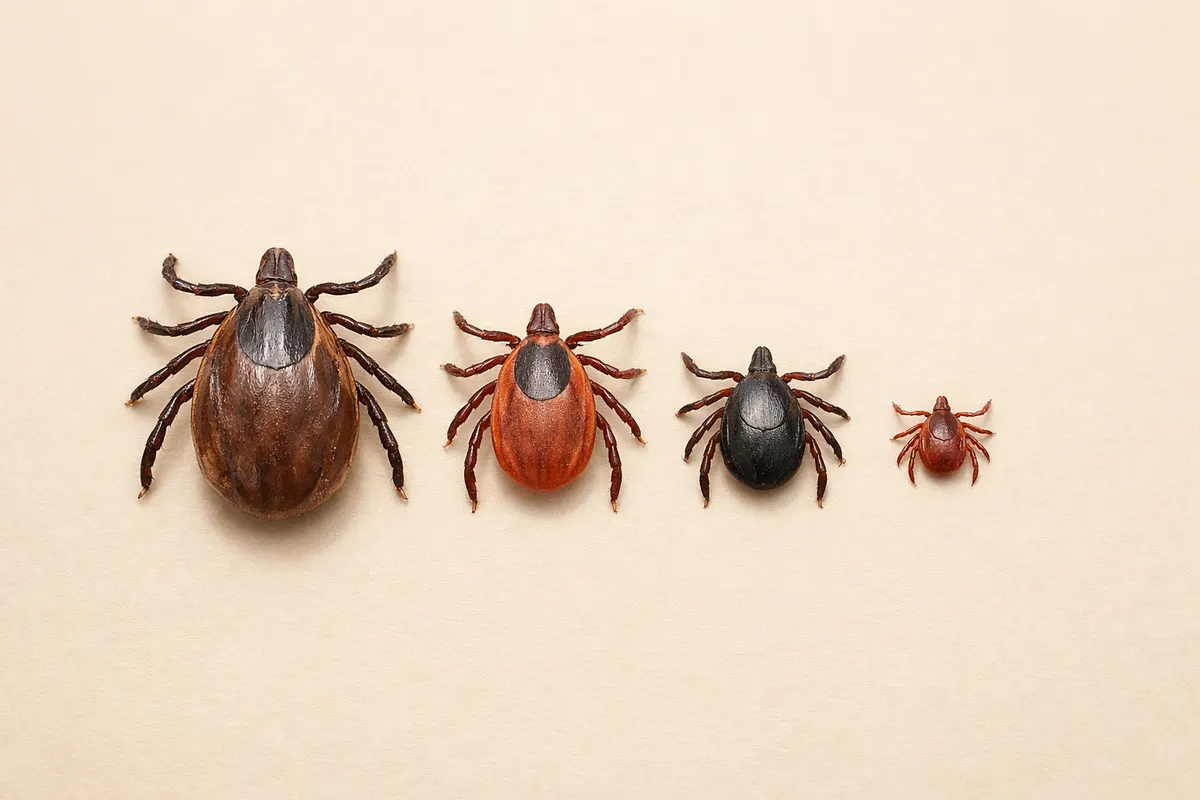
Quick answer: identifying tick species at a glance
Tick species are most reliably separated by a handful of visible traits: overall color and ornamentation, scutum (shield) patterns, mouthpart length and shape, presence of a single white spot, and body shape when engorged. Geography and the tick’s life stage (larva, nymph, adult) narrow possibilities further.
If you only need a fast, cautious read: a small, dark tick with long mouthparts in the northeastern U.S. is often a blacklegged (deer) tick; a reddish-brown tick with a single white spot on the back is commonly a lone star tick; a larger, patterned tick with festoons in grassy areas is often an American dog tick. Those are rules of thumb, not certainties.
Photos that include scale, a dorsal close-up, and an angle showing the mouthparts give the most power for identification. When a precise ID matters for exposure or treatment decisions, follow up with a trusted lab, local extension office, or health professional — a photo alone can be misleading.
- Key visual clues: scutum patterns, mouthpart length, ornamentation, festoons, and body color.
- Context matters: location, host species (human, dog), and season change the likely species.
- Photo-first approach: give a dorsal shot, a close-up of the head, and a full-body view with a ruler or coin for scale.
What we mean by tick species and why it matters
When people search for 'tick species' they usually want to know which type of tick they found and whether it changes the risk of tick-borne disease or the best next steps. 'Tick species' refers to the taxonomic category (for example Ixodes scapularis, Dermacentor variabilis, Amblyomma americanum) and the practical identification features that separate those taxa.
Different species have different geographic ranges, host preferences, and disease associations. For example, Ixodes scapularis (blacklegged or deer tick) is a primary vector for Lyme disease in much of the eastern U.S., while Amblyomma americanum (lone star tick) is linked to ehrlichiosis and certain allergic reactions. That said, species alone don’t tell you whether a particular tick carried a pathogen — only testing can do that.
Practically, identifying the species helps prioritize follow-up: whether to save the tick for testing, how long to watch for symptoms, or which local resources to contact. It also helps avoid common misidentifications with lookalikes like small beetles, chiggers, or immature mites.
- Species affects likely pathogens and therefore may change monitoring advice (not immediate clinical diagnosis).
- Host and season narrow likely species — a dog in the Midwest in summer gets different common ticks than a hiker in fall in New England.
- Identification informs whether to keep the specimen, photograph it, or pursue lab testing through local public health or veterinary channels.
Key clues to check when identifying tick species
Start with the big-picture features that are easiest to see in a photo: size (and whether the tick is engorged), overall color, and whether the tick’s back has an ornamented shield (scutum). Patterned scutums—often with bright or pale markings—point toward Dermacentor or Amblyomma species in many regions, while uniformly dark backs often indicate Ixodes species.
Mouthparts are decisive when visible. Long, slender mouthparts that extend forward beyond the body’s edge are typical of Ixodes (blacklegged) ticks. Short, stout mouthparts are more typical of Dermacentor (American dog tick) and Rhipicephalus (brown dog tick). A single conspicuous white spot near the center of the scutum is a classic sign of an adult female lone star tick (Amblyomma americanum).
Look for secondary features: festoons (small lobed segments along the posterior edge), the shape of the basis capituli (the base of the mouthparts), leg color, and presence or absence of an anal groove. For example, Ixodes species have an anal groove that arches in front of the anus on the underside—an observation that usually requires a ventral view or a high-quality close-up.
- Scutum: patterned vs. uniform; ornamentation suggests Dermacentor or Amblyomma.
- Mouthparts: long and forward-projecting (Ixodes) vs. short and stubby (Dermacentor/Rhipicephalus).
- White spot: lone star adults often have a single central white spot (females).
- Festoons: presence suggests Dermacentor or Rhipicephalus; absence is typical for Ixodes.
- Anal groove: visible under good magnification—arched in front for Ixodes species.
A step-by-step workflow to narrow down tick species
1) Capture context and scale. Photograph the tick beside a ruler, coin, or a metric scale. Note where and when it was found, what host it was on (human, dog, deer), and whether it was attached or crawling. Geographic region and season eliminate many options quickly.
2) Take three essential shots: a clear dorsal (top) view, a lateral or head-angle that shows the mouthparts, and an overall photo showing size relative to the scale object. If possible and safe, a ventral shot helps with features like the anal groove and basis capituli. Use soft diffuse light and a steady hand or tripod for macro clarity.
3) Compare major characters. Use the dorsal photo to examine scutum pattern and overall color; use the head-angle to judge mouthpart length. If you see a single bright spot on the dorsum, consider lone star; if the tick is small and dark with long mouthparts, Ixodes is likely; if it’s large with a patterned scutum and festoons, think Dermacentor or Amblyomma.
4) Cross-check life stage and engorgement. Nymphs and larvae are much smaller and sometimes look different from adults; engorgement changes color and hides ornamentation. Adjust expectations accordingly and avoid overconfident labeling when only immature stages or engorged specimens are available.
5) Decide on next actions. If the species and exposure raise concern, keep the specimen in a sealed container (dry, not in alcohol unless local guidance recommends it), photograph it well, and consult local public health or a veterinarian for testing instructions. If you prefer a fast first-pass interpretation, use photo-identification resources or a guided checklist before escalating to laboratory testing.
- Photo checklist: dorsal, head/mouthparts, ventral (if safe), and scale reference.
- Context checklist: exact location, host species, date, and activity when encountered.
- Decision checklist: low concern (tick removed cleanly, unlikely vector), moderate concern (species associated with local disease), or high concern (attachment duration long or symptoms present)—use this to choose further steps.
Examples: matching common photos to likely tick species
Example 1 — Small dark tick found on a hiker in New England in May: Dorsal photo shows a uniform dark brown back with no obvious ornamentation, and a close-up shows long mouthparts extending beyond the body margin. This pattern fits an Ixodes scapularis (blacklegged/deer tick) nymph or adult, depending on size. Because Ixodes is a primary Lyme vector in that region, note the attachment time and consult local guidance if attachment exceeded 36–48 hours.
Example 2 — Reddish-brown tick with a single white spot on the dorsal center found on a person in the Southeast in June: The distinctive central white spot on an otherwise reddish-brown scutum strongly suggests an adult female Amblyomma americanum (lone star tick). Lone star ticks can cause ehrlichiosis and have been associated with alpha-gal allergic reactions; consider follow-up if symptoms appear or the attachment was prolonged.
Example 3 — Large tick with patterned scutum and visible festoons found on a dog after yard play in the Midwest: A patterned shield and festoons are consistent with Dermacentor variabilis (American dog tick) or Amblyomma species. Because dogs commonly carry Dermacentor and Rhipicephalus in many areas, compare the mouthparts (shorter in Dermacentor) and consult your veterinarian about removal and monitoring.
Example 4 — Small, uniform tan tick found in a home with multiple ticks on a dog: Brown dog tick (Rhipicephalus sanguineus) often thrives indoors on dogs and shows a uniform reddish-brown color with short mouthparts. Multiple indoor ticks on pets suggest a need for veterinary advice and environmental checks, as brown dog ticks can complete life cycles indoors.
- Ixodes (blacklegged): small, dark, long mouthparts, northern/eastern U.S. common.
- Amblyomma americanum (lone star): reddish, female with central white spot, southeastern U.S.
- Dermacentor variabilis (American dog tick): larger, patterned scutum, festoons visible, common in grass.
- Rhipicephalus sanguineus (brown dog tick): uniform brown, short mouthparts, often found on dogs and indoors.
Limitations: when a photo is not enough for tick species ID
Photos have real utility but clear limits. Engorgement alters color and hides scutum ornamentation; immature life stages (larvae and nymphs) lack the same adult markings and are much harder to assign to species. Low-resolution images, poor lighting, and obstructed mouthparts frequently prevent a confident ID.
Geographic overlap and lookalikes further complicate matters. Some non-tick arthropods (mites, beetle larvae) can be mistaken for ticks in poor photos. Likewise, regional variations mean that the same species may look slightly different across ranges. When a precise species label affects medical or veterinary decisions, rely on an expert exam or a lab that accepts specimens for morphological or molecular testing.
Separate outcomes by confidence level: high-confidence IDs are possible when multiple diagnostic characters are visible (clear scutum pattern, visible mouthparts, and a clear ventral view); moderate-confidence IDs use fewer or partial clues; and low-confidence or uncertain outcomes occur when photos show only general size and color or when the tick is engorged or damaged. For moderate or low confidence, collect better photos, add context, or seek a professional check rather than acting on a single tentative label.
- High confidence: clear dorsal scutum pattern, mouthparts visible, and known location/host.
- Partial confidence: only dorsal view or only a size reference—useful for narrowing but not confirming.
- Uncertain: engorged specimens, larvae/nymphs without diagnostic marks, or blurred photos—seek verification.
- When necessary, preserve the tick following local public health guidance and contact testing or veterinary services for definitive identification.
Related guides
Need a fast first-pass check for a tick species?
After you collect clear photos and context, use Tick Detector - Tickzen tools or consult the Tick Identification Chart to get a guided first pass. Our identification chart at https://tick-detector.app/tools/tick-identification-chart walks through the same visual clues used here and helps you decide whether to keep the specimen, seek lab testing, or contact a vet or public health office.
Take the next step →Frequently asked questions
How can I tell the difference between a deer tick and a dog tick from a photo?
Deer ticks (Ixodes species) are usually smaller and darker with long mouthparts that extend past the front of the body; they often lack bright scutum ornamentation. Dog ticks (Dermacentor variabilis) are generally larger, often show a patterned scutum and have visible festoons at the rear. A clear dorsal photo plus a head-angle that shows mouthpart length helps distinguish them. If the tick is engorged, these differences become harder to see and identification confidence drops.
Does the presence of a lone star tick mean I'm at immediate risk for disease?
Finding a lone star tick (Amblyomma americanum) does not automatically mean you have been infected. Lone star ticks are associated with certain infections and allergic reactions, but risk depends on attachment duration, whether the tick fed, and local pathogen prevalence. Monitor for symptoms and consult local health guidance; when in doubt, photograph the tick, preserve it if recommended, and reach out to a public health or veterinary resource for next-step advice.
What photos should I take to help identify a tick species?
Take at least three photos: a clear dorsal (top) view showing the scutum, a close-up of the head/mouthparts from the front or side, and a full-body shot that includes a ruler or coin for scale. If safe, a ventral shot helps with features like the anal groove. Use natural diffuse light, steady the camera, and avoid touching the tick. Record the location, host species, and date — that context greatly increases identification accuracy.
When should I send a tick for laboratory identification or testing?
Consider laboratory identification or testing if the tick is of a species known to carry serious local pathogens, if it was attached for a long time (often >24–48 hours), if the exposed person or pet develops symptoms, or if your local health authority recommends testing. Testing policies vary by region and organism; contact local public health departments, extension services, or veterinary clinics for instructions on specimen handling and submission.